

|
|
|
|
|
|
||||||||||||
|
|
 |
|
|
|
"White Sheet" for PhysioFlow® The ability to quantify cardiac output and other hemodynamic indices easily and non-invasively has important applications for the routine clinical evaluation of patients with cardiovascular disease. These measurements have been made in the past using a direct method known as thermodilution. However, this method is cumbersome, invasive, expensive, and carries an inherent risk. PhysioFlow® is a device for estimating cardiovascular hemodynamics non-invasively based on the principle of thoracic electrical bioimpedance - essentially this technology quantifies a high frequency, low magnitude signal across the patient's chest. The technology is based on the concept that flow can be measured by electrical resistance of tissue to the flow of electrical current. Changes in bioimpedance, resulting from the changes in volume and velocity of blood in the aorta, are inversely related to stroke volume, allowing the estimation of cardiac output. The technique requires only the addition of 6 electrodes on specific locations across the thorax and neck. Based on this concept, both cardiac and systemic parameters can be estimated; these include heart rate, stroke volume, cardiac output, ejection fraction, contractility index, early diastolic function ratio, and systemic vascular resistance.
Applications The ability to measure cardiac output and other hemodynamic parameters non-invasively has many clinical applications. For example, a fundamental characteristic of patients with chronic heart failure is an impaired ability to increase cardiac output appropriately with exercise. The fact that many patients improve their functional capabilities after interventions such as cardiac resynchronization therapy, left ventricular assist device implantation, pharmacologic therapy, or exercise training is largely related to an improved ability to increase cardiac output with exercise. Quantifying cardiac output can therefore provide important information about these and other interventions. Recent studies have also demonstrated that this information is valuable in predicting outcomes in patients with cardiovascular disease.
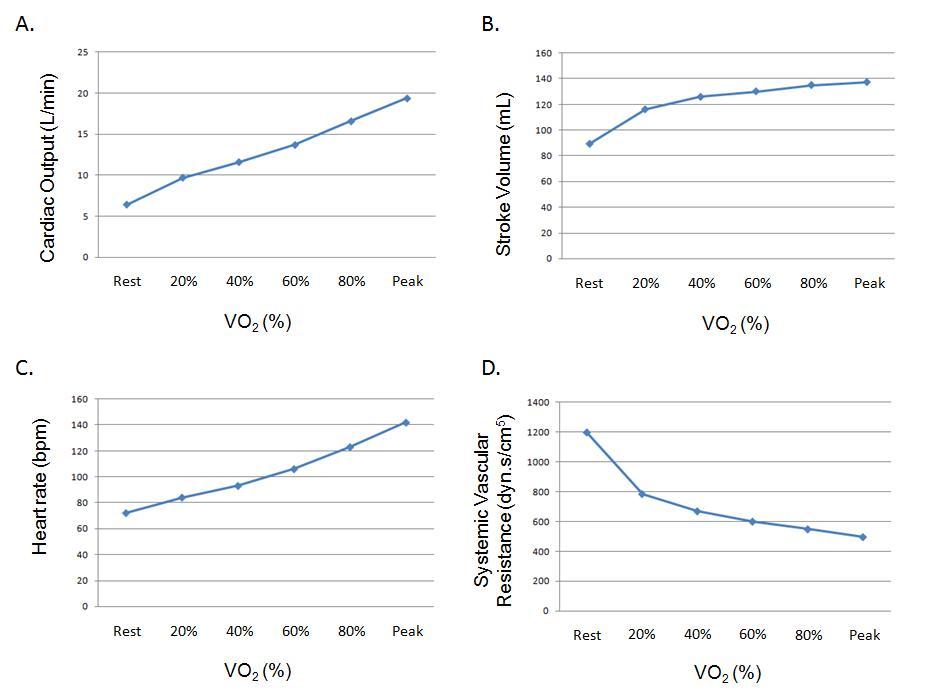
In one VA medical center, cardiopulmonary exercise test responses and PhysioFlow® non-invasive hemodynamic monitoring data were measured at exercise times corresponding to 20%, 40%, 60%, 80% and 100% of peak oxygen uptake. Below, hemodynamic data from a patient referred for exercise testing for clinical reasons are plotted as a percentage of peak VO2. Note that this patient exhibits a relatively normal response, and was found to have normal ventricular function by echocardiography, a good exercise capacity, and no significant ECG abnormalities during exercise.
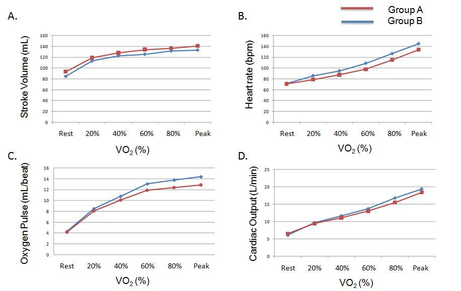
Similarly, PhysioFlow® non-invasive hemodynamic monitoring responses can be compared between groups or within groups following an intervention. Below, a group of patients with abdominal aortic aneurysm disease (Group A) is compared to a group of patients referred for exercise testing for clinical reasons (Group B) at matched percentages of peak VO2. Expressing the data as a relative percentage permits a comparison between groups that is independent of maximal exercise capacity. While both groups exhibit relatively normal responses and are similar to one another, the ability to measure these hemodynamic responses non-invasively and to compare them provides important information about various disease states as well as interventions in patients with cardiovascular disease.
Jonathan Myers, Ph.D |
|
|
|
|
This site and all contents are © Copyright 2009 - All Rights Reserved by NeuMeDx, Inc., Bristol, PA 19007
| Site Map |